New Machine Learning Model Could Accurately Detect Breast Cancer

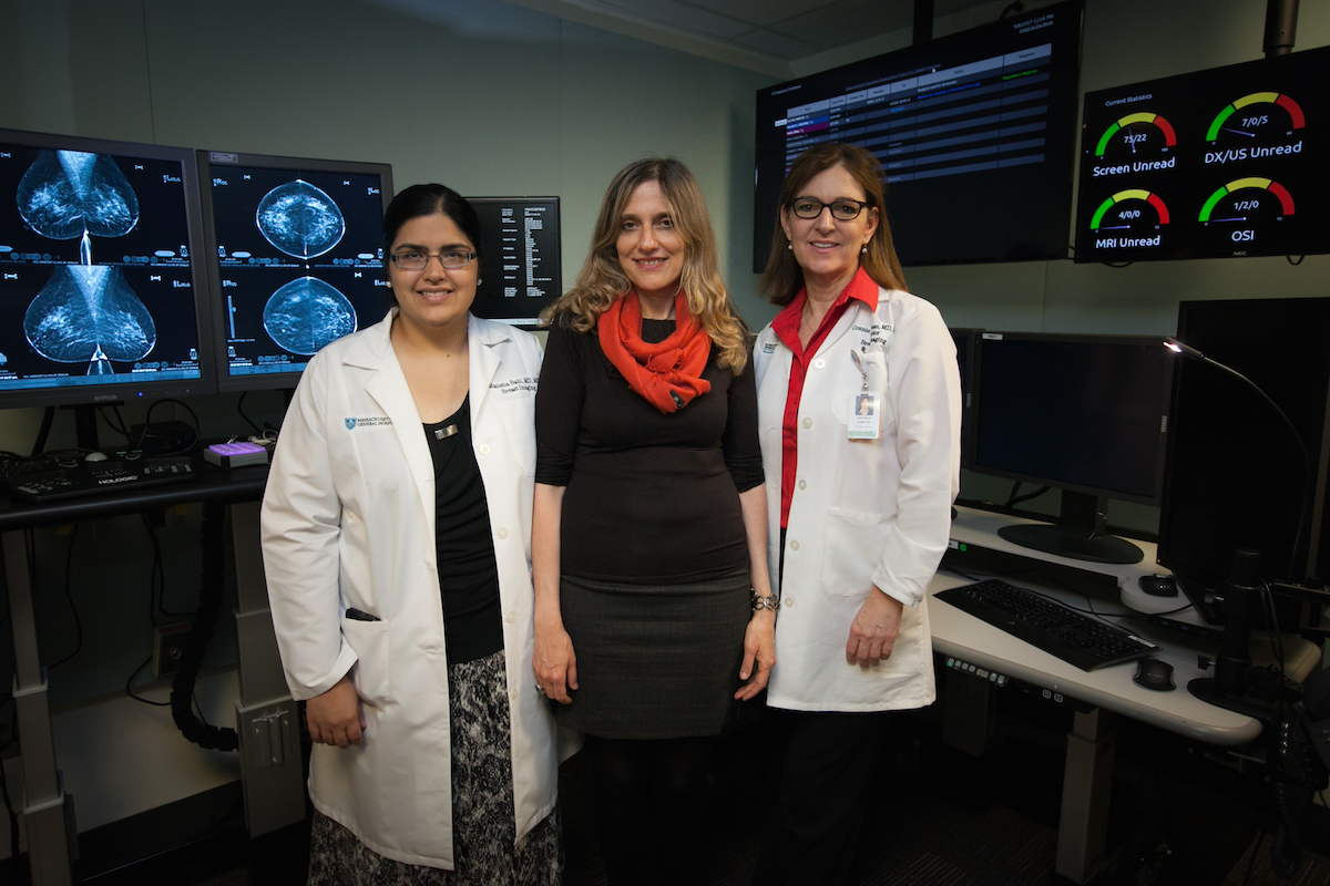
MIT professor Regina Barzilay (center) with Mass General collaborators Manisha Bahl and Constance Lehman. / Photo provided
Harnessing the power of machine learning, Boston researchers hope to change breast cancer detection techniques for the better.
Scientists from Massachusetts General Hospital and MIT’s Computer Science and Artificial Intelligence Laboratory (CSAIL) believe their new method can accurately screen high-risk lesions for breast cancer without requiring surgery to remove them. Machine learning lets artificial intelligence systems learn and improve on their own—without being explicitly programmed—once they’re given information to analyze.
Other methods only allow for doctors to determine if a lesion is cancerous during surgery. Ninety percent of the time, the lesions are non-cancerous—making an invasive and expensive procedure totally unnecessary.
The new model can detect cancer based on patient information, using a machine learning method called a random-forest classifier. In this method, researchers fed information on over 600 high-risk lesions—including details about the patient’s age and race, family history, past biopsies, and pathology reports—to an artificial intelligence. In trials, that artificial intelligence was able to interpret that data so effectively that the method resulted in fewer unnecessary surgeries and specifically diagnosed more cancerous lesions.
The model identified 97 percent of cancers, compared to 79 percent rate possible in existing methods, according to an MIT statement. Notably, it also reduced the number of unnecessary surgeries by more than 30 percent compared to current methods.
“Because diagnostic tools are so inexact, there is an understandable tendency for doctors to over-screen for breast cancer,” says lead author Regina Barzilay, a professor at MIT CSAIL, in the statement. “When there’s this much uncertainty in data, machine learning is exactly the tool that we need to improve detection and prevent over-treatment.”
For Barzilay, the project is personal. She was diagnosed with breast cancer in 2014 and is now in remission. When she looked at her past mammograms, she noticed that her tumor had gone undetected for two years. With this new model, Barzilay hopes to take away some of that uncertainty.
Barzilay co-wrote the new paper with Constance Lehman, a professor at Harvard Medical School and chief of the Breast Imaging Division at MGH’s Department of Radiology, as well as Mass General’s Manisha Bahl and CSAIL graduate students Nicholas Locascio, Adam Yedidia, and Lili Yu. It was published Tuesday in Radiology.
Currently, doctors rely on biopsies to investigate whether a lesion is cancerous. Around 70 percent are benign, 20 percent are malignant, and 10 percent are high-risk lesions, according to the MIT statement.
Some doctors surgically remove high-risk lesions in all cases. Others perform surgery for specific lesions with higher cancer rates, but can miss other cancerous lesions.
“In the past, we might have recommended that all high-risk lesions be surgically excised,” Lehman says in the MIT statement. “But now, if the model determines that the lesion has a very low chance of being cancerous in a specific patient, we can have a more informed discussion with our patient about her options. It may be reasonable for some patients to have their lesions followed with imaging, rather than surgically excised.”
In the coming months, researchers will test similar models to see how well the same approach works with other forms of cancer and even other diseases. But it’s also something that will have immediate, real-world applications: Radiologists at Mass General will now incorporate it into their treatment of breast cancer patients.
The team is still working to hone their method. As is common with machine learning models, theirs will only improve the more information it’s given to work with.
“In future work, we hope to incorporate the actual images from the mammograms and images of the pathology slides, as well as more extensive patient information from medical records,” says Bahl.


