Doctors Are Using 3-D Printed Models to Prep for Face Transplants

Example of a 3D-printed model used in preparing for face transplant surgery. Photo provided to bostonmagazine.com
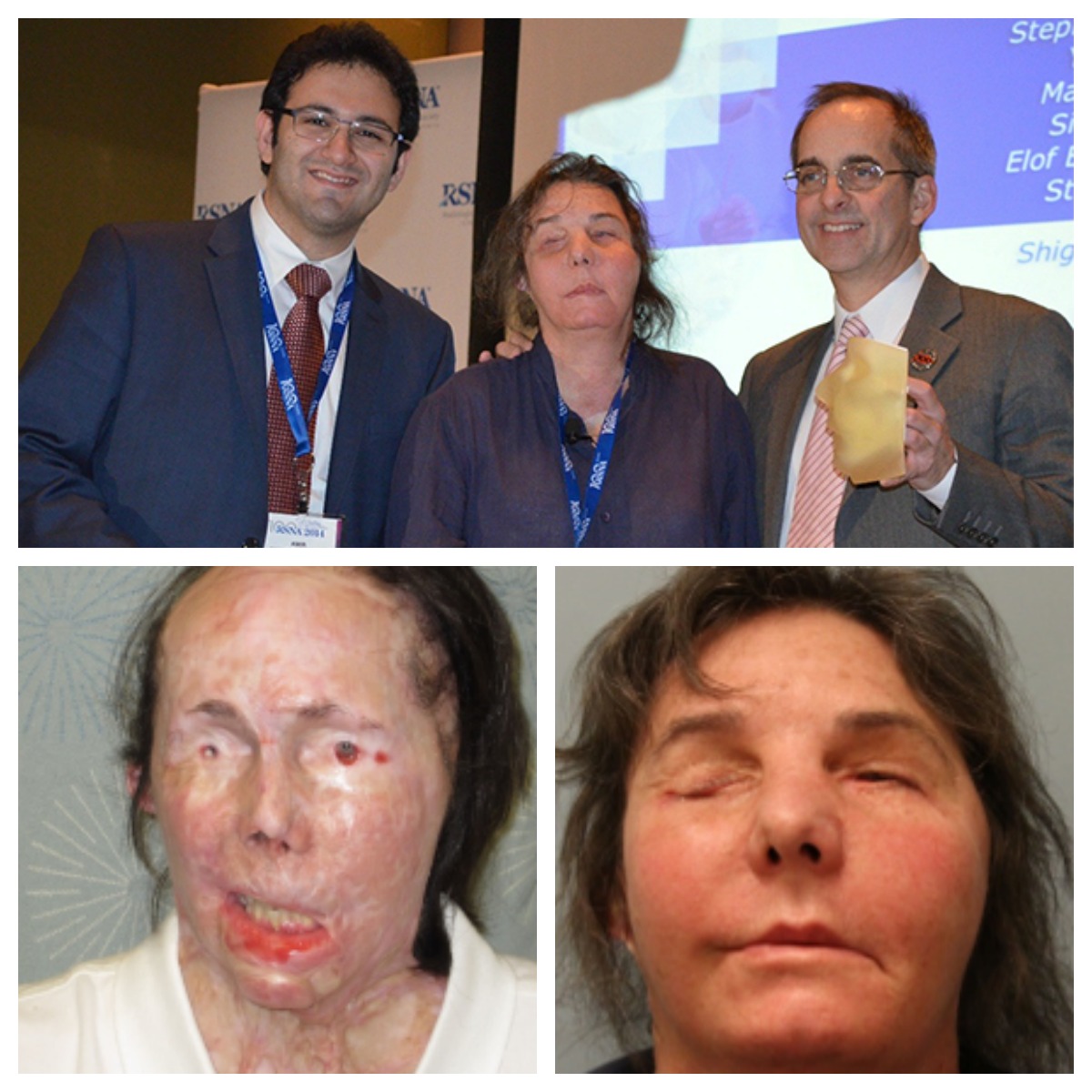
Carmen Tarleton remembers the moment she saw her new face for the first time.
After being attacked in 2007 by her now ex-husband, Tarleton suffered a severe chemical burn covering 80 percent of her body, including her head and face. The injury left her with only a partial nose and lip as well as significant scarring on her neck, which prevented her from holding her head up. After nearly 60 surgeries, the Vermont resident ultimately received a face transplant at Brigham and Women’s Hospital on Valentine’s Day 2013.
“I expected it to be this big, dramatic thing but when I looked into the mirror that first time, I just had this sense of relief,” she says. “All the scarring in my neck was gone when I woke up from surgery. I had a complete neck, and that’s what I needed.”
Doctors at the Brigham have been using 3-D printed models of bones for some time now, in order to help them plan and prepare for face transplant surgeries like Tarleton’s. Frank Rybicki, the director of the hospital’s Applied Imaging Science Laboratory, says that the models—created from CT images of the recipient’s skull—are used to determine whether the anatomy and bone structure of the donor and recipient are compatible. Now, thanks to advances in 3-D printing technology, doctors are able to look at another crucial element: soft tissue.
“There’s no better way to understand the anatomy you’re operating on than to hold it in your hand,” Rybicki says. “We used to be able to just print bones, but now a whole new set of technologies are allowing us to simulate tissue types like blood vessels and skin. It’s a much better medical model than we’ve ever had before.”
Rybicki says that one of the benefits of using 3-D printed models of bone and tissue is that it gives surgeons an opportunity to practice technique before the actual operation. This leads to more accurate results as well as shorter operating times—a typical face transplant surgery can take 20 hours. The models are also useful in monitoring a patient’s progress once the surgery is complete, he adds.
“Before 3-D printing, the only way to monitor soft tissue changes over time was to take photographs or to use the CT images to make simplistic measurements,” he says. “[These] models allow the soft tissues to be rendered exactly as they are in the patient so that these changes can be systematically followed and studied.”
To date, there have been seven face transplant surgeries performed at the Brigham. Tarleton, who was the fourth surgery of the seven, says that though her recovery has been long and painful, the transplant surgery was “heaven-sent.”
“The transplant has had so many blessings for me, and my quality of life on a daily level has improved greatly,” she says, adding that she is no longer on narcotics for the pain. “It’s been more than seven years since my initial injury, and I just keep getting better and better.”
Photos provided.